|
|
|
|
|
|
|
Flicker PerimetryChris A. Johnson, Ph.D. INTRODUCTIONThe ability to detect intermittent light and dark alternations of a visual stimulus (flicker or temporal visual processing) is an important component of visual function throughout the field of view. Rapid changes in the luminance or contrast of a stimulus can be important for detecting environmental changes, motion, and awareness of objects in peripheral vision. A thorough description of the variables influencing and mechanisms underlying flicker sensitivity is beyond the scope of this presentation, but there are several references that can provide a comprehensive review.1-5 Flicker sensitivity has been a topic of interest to many investigators for nearly 200 years. For many years, psychophysical flicker sensitivity has been reported to be diminished in glaucoma and ocular hypertension.6 Tyler reported that glaucoma patients demonstrated a high temporal frequency flicker sensitivity loss, and up to 90% of ocular hypertensives also exhibited a high temporal frequency deficit.6 However, the ocular hypertensive cases were actually the fellow eyes of patients who had glaucoma in the other eye, which draws into question the likelihood of disease in the fellow eyes. Additionally, subsequent investigations have reported that reductions in sensitivity with age are more prominent for high temporal frequencies than for low and intermediate temporal frequencies.5 Thus, the high temporal frequency deficit may be related to the normal aging process rather than a selective reduction in high frequency flicker sensitivity that is related to glaucoma, and this has been confirmed in subsequent studies.7,8 Nonetheless, it is important to recognize that the ability to detect flicker is a sensitive and early indicator of functional loss in glaucoma, and subsequent studies have confirmed this result. Flicker perimetry is a visual field test procedure that evaluates an observer’s ability to detect light/dark stimulus alternations (flicker) at various locations in the field of view. In general, there are three types of flicker perimetry test procedures that have been utilized: (1) contrast modulation flicker, (2) critical flicker fusion (CFF), and (3) luminance pedestal flicker. Contrast modulation flicker uses a stimulus that is matched in luminance to the background. The contrast of the stimulus is then modulated temporally according to a fixed frequency, and the amplitude of flicker modulation needed for detection of the stimulus is determined (Figure 1a) for different rates of flicker. Critical flicker fusion (CFF) uses a sinusoidal grating with 100% (or close to 100%) contrast, and determines the maximum frequency or rate of flicker that can be distinguished from a steady, uniform field (Fig 1b). Luminance pedestal flicker presents a flickering stimulus superimposed on a pedestal of steady light and determines the amount of flicker that is needed to distinguish the flicker from a steady uniform stimulus (Figure 1c). There are advantages and disadvantages of each procedure, and a brief description of findings for each procedure is presented below. Only in a few instances have different methods of measuring flicker sensitivity thresholds been compared.9 Methods other than the ones described above have also been used in characterizing flicker perimetry.10-12
Contrast Modulation Flicker
Critical Flicker FusionCritical flicker fusion perimetry (CFF) determines the highest flicker frequency that can be distinguished from a uniform steady stimulus. Typically a fixed high contrast modulation is employed (near 100% contrast). CFF perimetry is best performed if the flicker modulation is about the average luminance of the background adaptation level. Several investigators have reported that this form of perimetry is superior to standard automated perimetry in its ability to detect glaucomatous visual field loss and evaluate the extent of glaucomatous visual field damage.13-19 Additionally, it has been reported that this form of flicker testing demonstrates minimal aging effects and is robust to a variety of factors (e.g., blur) that have traditionally been difficult for many other forms of perimetry. 13-15 Luminance Pedestal FlickerMany of the automated perimeters in use today have a white hemispherical bowl that serves as the uniform adaptation background and/or use light emitting diodes (LEDs) as the stimulus light source. In these instances, it is difficult to have a flicker stimulus that is identical in luminance and chromaticity to the background. One solution to this problem has been the development of a procedure in which a flickering stimulus is superimposed on a luminance increment, and it is known as luminance pedestal flicker.20-23 The observer’s task is to determine whether the luminance increment is flickering or is steady by pressing a response button when flicker is detected. To date, only preliminary investigations of this procedure in a perimetric context has been accomplished,22 and evaluations of patients with ocular and neurologic disorders has not been attempted with this technique. Future assessments of the clinical utility of this procedure will be of great interest to many practitioners and may yield additional information that will be of assistance for diagnostic purposes. One of the difficulties
associated with the use of different methods of performing flicker perimetry
concerns the selection of the most appropriate procedure for clinical
diagnostic purposes. Although a full
comparison of all procedures has not been performed to date, a direct
investigation of contrast modulation flicker and critical flicker fusion
perimetry has been performed in a group of participants with normal visual
function and a group of patients with glaucomatous visual field loss.9
A Receiver Operating Characteristic (ROC)
analysis was performed for both procedures to determine their sensitivity
(ability to detect damage due to glaucoma) and specificity (ability to
correctly classify individuals with normal visual function and no evidence of
glaucoma) for a large variety of different decision rules. It was also possible to perform both test
procedures using the same prototype automated flicker perimeter. In this manner, it was possible to evaluate
the clinical efficacy of each procedure in a manner that would permit them to
be directly compared. The figures to
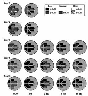
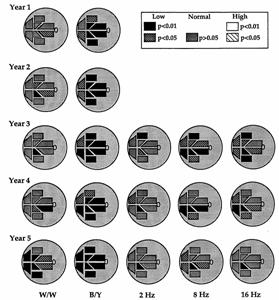
As with most clinical diagnostic test procedures, each of the methods of performing flicker perimetry has advantages and disadvantages that must be considered. TMP is more sensitive for detecting early glaucomatous visual field loss, but the selection of appropriate target size, background luminance, stimulus onset-offset conditions, test strategy, and other properties of the test procedure are highly important. For example, by using an intermediate temporal frequency that is near the peak of the temporal contrast sensitivity function, test retest variability can be minimized, and the dynamic range of the procedure can be maximized. CFF perimetry can provide very useful information about the upper temporal frequency limits of processing flicker information, although the variability may be higher and the dynamic range may be limited. Additionally, CFF perimetry can be an easier procedure to implement on existing instrumentation, although its use on video monitor display systems would limit the possible temporal frequencies that could be displayed. LPF perimetry is a procedure that can be the most easily implemented on existing commercial perimetric instruments. This makes its availability a desirable characteristic. However, the stimulus presentation consists of both a luminance onset (pedestal) and the initiation of a flickering stimulus superimposed on the luminance pedestal. For some subjects, there may be confusion and errors produced when subjects are asked to respond only to the flicker and not the stimulus onset. Evaluation of the influence of basic stimulus and test parameters on flicker sensitivity has been examined by many investigators, along with procedures to optimize the methodology for clinical testing of patients.1-23 These investigations are important because small variations in pupil size, adaptation level and may other features can dramatically alter flicker sensitivity. It is critical to apply test procedures that are robust to non-pathologic influences on flicker sensitivity, and to implement test procedures that are best designed to provide stable, reproducible test results. In view of the many stimulus parameters that can influence the sensitivity to flicker, this represents a challenging and formidable task. However, many investigators have been able to accomplish this goal in recent years. From a clinical perspective, flicker perimetry in its various forms has been reported to be a sensitive indicator of early functional damage for a variety of disorders, including age-related macular degeneration and retinal diseases,26-32 glaucoma, 6-10,13-19,24,25 and other ocular and neurologic disorders. The figures below present examples of visual field progression in the right eye of two patients with glaucomatous visual field loss. Results are shown schematically for nerve fiber bundle regions in the superior and inferior visual field for normal (p> 0.05), and upper (high) and lower (low) 95 and 99% confidence limits, designated as p<0.05 and p<0.01, respectively. Five years of results are presented for standard automated perimetry (W/W) and Short Wavelength Automated Perimetry (B/Y) and three years of results are presented for TMP perimetry obtained for flicker rates of 2, 8 and 16 Hertz. Note that the deficits for TMP perimetry (especially at 8 Hz) are predictive of future visual field loss for standard automated perimetry.
Additionally, recent techniques have been developed that utilize flicker perimetry as a method of testing the peripheral visual field of young infants.11,12 The use of flicker as a stimulus for evaluation of the peripheral visual field of infants is particularly appropriate because it is one of a few stimuli that an infant can attend to for prolonged periods of time. In this manner, important visual field information can be obtained from this young population. In summary, flicker perimetry in all of its forms has made it possible to evaluate peripheral visual function in an efficient manner, provides greater sensitivity for detecting early pathologic changes, and provides the opportunity to evaluate the visual field of individuals that otherwise may not be assessable. References1.
Kelly
DH: Flicker. In Handbook of Sensory Physiology, Vol VII/4 (L Hurvich and D
Jameson, eds), Chapter 11, 2.
McKendrick
AM, 3. Kelly DH: Visual responses to time-dependent stimuli: 1. Amplitude sensitivity measurements. J Opt Soc Am, 1961, 51: 422-429. 4. Kim CB, Mayer MJ: Foveal flicker sensitivity in healthy aging eyes. II. Cross-sectional aging trends from 18 through 77 years of age. J Opt Soc Am, 1994, 11: 1958-1969. 5.
Casson EJ, 6.
7.
Casson
EJ and 8.
Casson
EJ, 9.
Yoshiyama
K, 10. Horn FK, Jonas JB, Korth M, Junemann A, Grundler A: The full-field flicker test in early diagnosis of chronic open-angle glaucoma. Am J Ophthalmol, 1997, 123: 313-319. 11. Delaney SM, Dobson V, Mohan KM, Harvey EM: The effect of flicker rate on measured visual field extent in very young children. Optom Vis Sci, 2001, 78: 846-852. 12. Delaney SM, Dobson V, Mohan KM: Measured visual field extent varies with peripheral stimulus flicker rate in very young children. Optom Vis Sci, 2005, 82: 800-806. 13. Lachenmayr BJ, Kojetinsky S, Ostermaier N, Angstwurm K, Vivell PM, Schaumberger M: The different effects of aging on normal sensitivity in flicker and light-sense perimetry, Invest Ophthalmol Vis Sci, 1994, 35: 2741-2748. 14. Lachenmayr BJ: The role of temporal threshold criteria in psychophysical testing in glaucoma. Curr Opin Ophthalmol, 1994, 5: 58-63. 15. Lachenmayr BJ, Gleissner M: Flicker perimetry resists retinal image degradation. Invest Ophthalmol Vis Sci, 1992, 33: 3539-3542. 16. Lachenmayr BJ, Drance SM: Diffuse field loss and central visual function in glaucoma. Ger J Ophthalmol, 1992, 1: 67-73. 17. Lachenmayr BJ, Drance SM, Airaksinen PJ: Diffuse field loss and diffuse retinal nerve fiber loss in glaucoma. Ger J Ophthalmol, 1992, 1: 22-25. 18. Lachenmayr
BJ, Drance SM, 19. Lachenmayr BJ, Drance SM, Douglas GR, Mikelberg FS: Light-sense, flicker and resolution perimetry in glaucoma: a comparative study. Graefes Arch Clin Exp Ophthalmol, 1991, 229: 246-251. 20. Anderson AJ, Vingrys AJ: Interactions between flicker thresholds and luminance pedestals. Vision Res, 2000, 40: 2579-2588. 21. Anderson AJ, Vingrys AJ: Multiple processes mediate flicker sensitivity. Vision Res, 2001, 41: 2449-2455. 22. Anderson AJ, Vingrys AJ: Effect of eccentricity on luminance-pedestal flicker thresholds. Vision Res, 2002, 42: 1149-1156. 23. Anderson AJ, Vingrys AJ: Effect of stimulus duration in flicker perimetry. Clin Experiment Ophthalmol, 2000, 28: 223-226. 24. Matsumoto C, Takada S, Okuyama S, Arimura E, Hashimoto S, Shimomura Y: Automated flicker perimetry in glaucoma using Octopus 311; a comparative study with the Humphrey Matrix. Acta Ophthalmol Scand, 2006, 84: 210-215. 25. Austin MW,
O’Brien CJ, 26. Stavrou EP, Wood JM: Central visual field changes using flicker perimetry in type 2 diabetes mellitus. Acta Ophthalmol Scand, 2005, 83: 574-580. 27. Phipps JA, Dang TM, Vingrys AJ, Guymer RH: Flicker perimetry losses in age-related macular degeneration. Invest Ophthalmol Vis Sci, 2004, 45: 3355-3360. 28. Phipps JA, Guymer RH, Vingrys AJ: Temporal sensitivity deficits in patients with high-risk drusen. Aust NZ J Ophthalmol, 1999, 27: 265-267. 29. Vingrys AJ, Pseudovs K: Localized scotomata detected with temporal modulation perimetry in central serous chorioretinoapthy. Aust NZ J Ophthalmol, 1999, 27: 109-116. 30. Mayer MJ, Spiegler SJ, Ward B. Glues A, Kim CB: Mid-frequency loss of foveal flicker sensitivity in early stages of age-related maculopathy. Invest Ophthalmol Vis Sci, 1992, 33: 3136-3142. 31. Mayer MJ, Spiegler SJ, Ward B. Glues A, Kim CB: Foveal flicker sensitivity discriminates ARM-risk from healthy eyes. Invest Ophthalmol Vis Sci, 1992, 33: 3143-3149. 32. Mayer MJ, Ward B, Klein R, Talcott JB, Dougherty RF, Glues A: Flicekr sensitivity and fundus appearance in pre-exudative age-related maculopathy. Invest Ophthalmol Vis Sci, 1994, 35: 1138-1149. Return to Computer Graphics Perimetry and other new stimuli: 1980 and beyond Return to Table of Contents
Copyright 2008. Imaging and Perimetry Society |
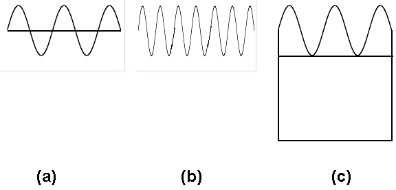
 As
described earlier, contrast modulation flicker perimetry is performed by using
a stimulus that is matched in luminance and color to the uniform background
which then undergoes a light and dark alternation (flicker) at a predetermined
frequency. The amplitude or contrast (modulation)
of flicker needed to detect the stimulus is then determined at key locations in
the visual field to yield a perimetric map of flicker sensitivity.
As
described earlier, contrast modulation flicker perimetry is performed by using
a stimulus that is matched in luminance and color to the uniform background
which then undergoes a light and dark alternation (flicker) at a predetermined
frequency. The amplitude or contrast (modulation)
of flicker needed to detect the stimulus is then determined at key locations in
the visual field to yield a perimetric map of flicker sensitivity.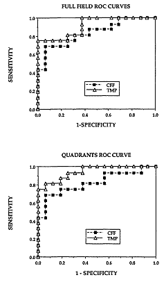 the right indicate the ROC
curves for flicker perimetry conducted using flicker modulation (temporal
modulation perimetry or TMP) and critical flicker fusion (CFF) as the response
measure. For the average performance of
the full field (top graph), the curve is higher for TMP than for CFF, but this
difference is even greater when the average performance of visual field
quadrants is examined (lower graph).
the right indicate the ROC
curves for flicker perimetry conducted using flicker modulation (temporal
modulation perimetry or TMP) and critical flicker fusion (CFF) as the response
measure. For the average performance of
the full field (top graph), the curve is higher for TMP than for CFF, but this
difference is even greater when the average performance of visual field
quadrants is examined (lower graph).