

Axenfeld-Rieger syndrome
Contributor: Jesse Vislisel, MD
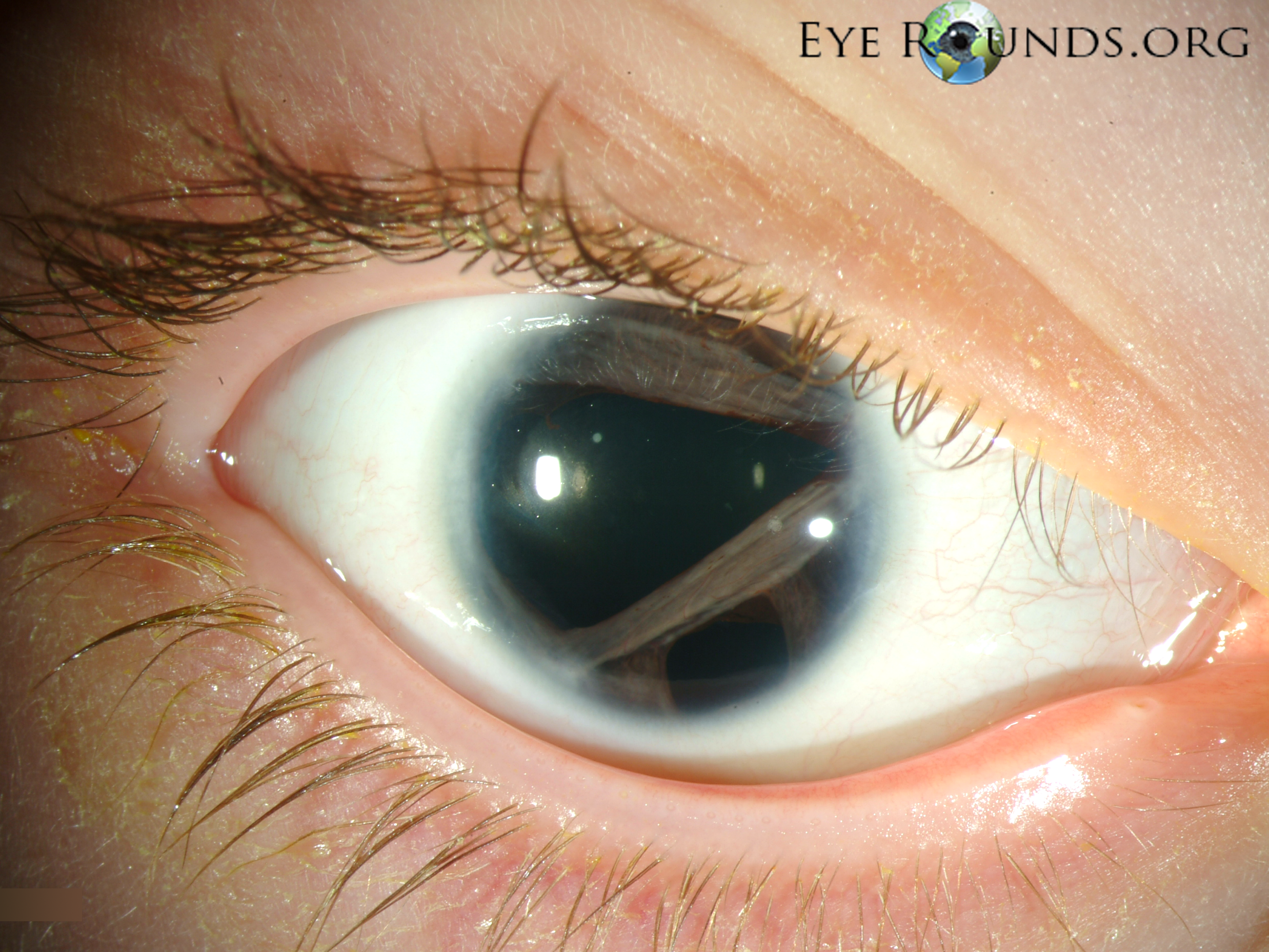
Corectopia (displacement of the pupil), polycoria (multiple holes in the iris), and posterior embryotoxon in a young boy with Axenfeld-Rieger syndrome.
(click on image for higher resolution image)
The patient also has glaucoma, redundant periumbilical skin, maxillary hypoplasia, and teeth anomalies. Posterior embryotoxon is a prominent white line running parallel to the limbus on the endothelial surface of the peripheral cornea. It is present in nearly all patients with Axenfeld-Rieger syndrome, but can also be seen in normal individuals.
OMIM #180500
Reference
Alward, W. L. M. Axenfeld-Rieger syndrome in the age of molecular genetics. Am. J. Ophthal. 130: 107-115, 2000. [PubMed: 11004268])

Ophthalmic Atlas Images by EyeRounds.org, The University of Iowa are licensed under a Creative Commons Attribution-NonCommercial-NoDerivs 3.0 Unported License.