

EyeRounds Online Atlas of Ophthalmology
Contributor: William Charles Caccamise, Sr, MD, Retired Clinical Assistant Professor of Ophthalmology, University of Rochester School of Medicine and Dentistry
*Dr. Caccamise has very generously shared his images of patients taken while operating during the "eye season" in rural India as well as those from his private practice during the 1960's and 1970's. Many of his images are significant for their historical perspective and for techniques and conditions seen in settings in undeveloped areas.
Category: Cornea
Corneal leukoma with iridodialysis
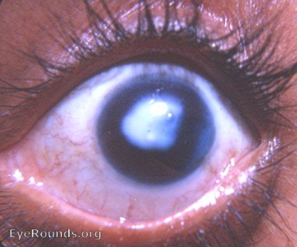
The leukoma is obvious. The pupil shows a flattening of its margin from 2 to 4 o'clock - the pupil margin is a straight line in this zone. This is pathognomonic of an iridodialysis. Careful examination will reveal the iridodialysis from 2 to 4 o'clock between the torn edge of the iris and the limbus - the area is slightly blue.

Ophthalmic Atlas Images by EyeRounds.org, The University of Iowa are licensed under a Creative Commons Attribution-NonCommercial-NoDerivs 3.0 Unported License.