

EyeRounds Online Atlas of Ophthalmology
Contributor: William Charles Caccamise, Sr, MD, Retired Clinical Assistant Professor of Ophthalmology, University of Rochester School of Medicine and Dentistry
*Dr. Caccamise has very generously shared his images of patients taken while operating during the "eye season" in rural India as well as those from his private practice during the 1960's and 1970's. Many of his images are significant for their historical perspective and for techniques and conditions seen in settings in undeveloped areas.
Category: Neuro-ophthalmology
Heterochromia with Horner's syndrome. Normal left eye is brown
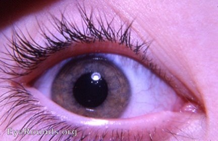
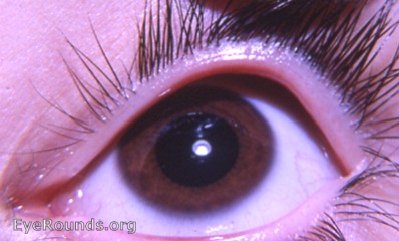
Patient has Horner's syndrome wih heterochromia iridis. The left eye is a normal brown. The Horner's right eye demonstrates a much lighter iris. In the binocular photo the lighter character of the right iris becomes definite at a zoom of 200% to 300%. The pupil seize difference in the two eyes is not evident because of cycloplegia from refracting drops. Heterochromia with Horner's syndrome appears only in those children who have acquired the syndrome at an early age - before the age of 2 years.Heterochromia iridis is an addition to miosis, ptosis, and anhidrosis in Horner's syndrome.
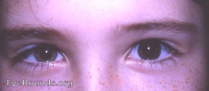
Binocular photo will reveal heterochromia at higher zoom values. Right eye is heterochromic.

Ophthalmic Atlas Images by EyeRounds.org, The University of Iowa are licensed under a Creative Commons Attribution-NonCommercial-NoDerivs 3.0 Unported License.