

EyeRounds Online Atlas of Ophthalmology
Contributor: William Charles Caccamise, Sr, MD, Retired Clinical Assistant Professor of Ophthalmology, University of Rochester School of Medicine and Dentistry
*Dr. Caccamise has very generously shared his images of patients taken while operating during the "eye season" in rural India as well as those from his private practice during the 1960's and 1970's. Many of his images are significant for their historical perspective and for techniques and conditions seen in settings in undeveloped areas.
Category: Vascular
Orbital varices, left eye
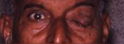
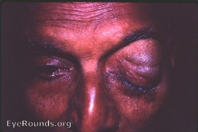
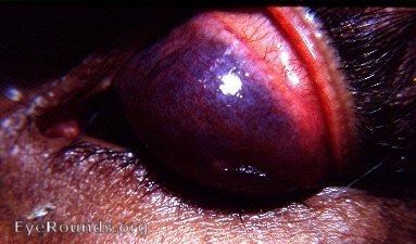
This Indian male was examined periodically from 1951 through 1968 without any change in the findings: on presenting himself in the clinic, he appeared to be normal. However, after bending over for 1 minute, he demonstrated marked protrusion of the lids of the left ye. Retraction of the upper lid revealed a huge bulging dark purplish lesion. Over a period of many minutes the lesion receded until the eye returned to a normal appearance.
The diagnosis was orbital varices.
The sequential photographs are: 1. the patient's normal appearance on walking into the Eye Clinic, 2. the patient's appearance in the upright position after having bent over for a few minutes, 3. orbital vascular abnormality apparent with retraction of the upper lid.
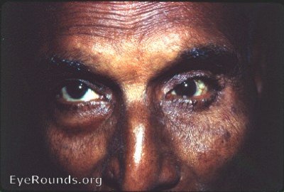
The patient presented with this relatively normal appearance in spite of huge orbital varices. (above)
Appearance after bending over (below)
Below, orbital vascular abnormality apparent with retraction of the upper lid.

Ophthalmic Atlas Images by EyeRounds.org, The University of Iowa are licensed under a Creative Commons Attribution-NonCommercial-NoDerivs 3.0 Unported License.