

Macular Hole, Full-Thickness, Extrafoveal/Eccentric
Contributor: Eric Chin, MD
Photographer: Cindy Montague, CRA
Patient
A 66-year-old female presented for a second opinion regarding a peripheral field defect in her left eye. She underwent epiretinal membrane (ERM) peeling at an outside office four months prior. Per records, this included internal limiting membrane (ILM) peeling and was complicated by post-operative vitreous hemorrhage. She says her vision improved some, however she had a large nasal field defect following this. She also had a history of glaucoma and had underwent prior selective laser trabeculoplasty (SLT) OU. She had no other complaints.
- BCVA cc: OD 20/20; OS 20/20
- IOP: OD 13, OS 15
- SLE: OD 1+ NS; OS 2+ NS, 1+ PSC
Fig 1: Fundus photograph OS: peripapillary atrophy; moderate to advanced optic nerve cupping; normal vessels; pigmentary changes inferotemporal to disc and superotemporal to the fovea center; there is a full-thickness macular hole about 1 disc diameter superior to the fovea center.

Fig 2: Fundus photograph OS: higher magnification image showing the eccentric/extrafoveal full-thickness macular hole (white arrows)

Fig 3-8: OCT OS: scant ERM remnants nasal to the fovea; thinning temporal in areas of prior membrane peeling.
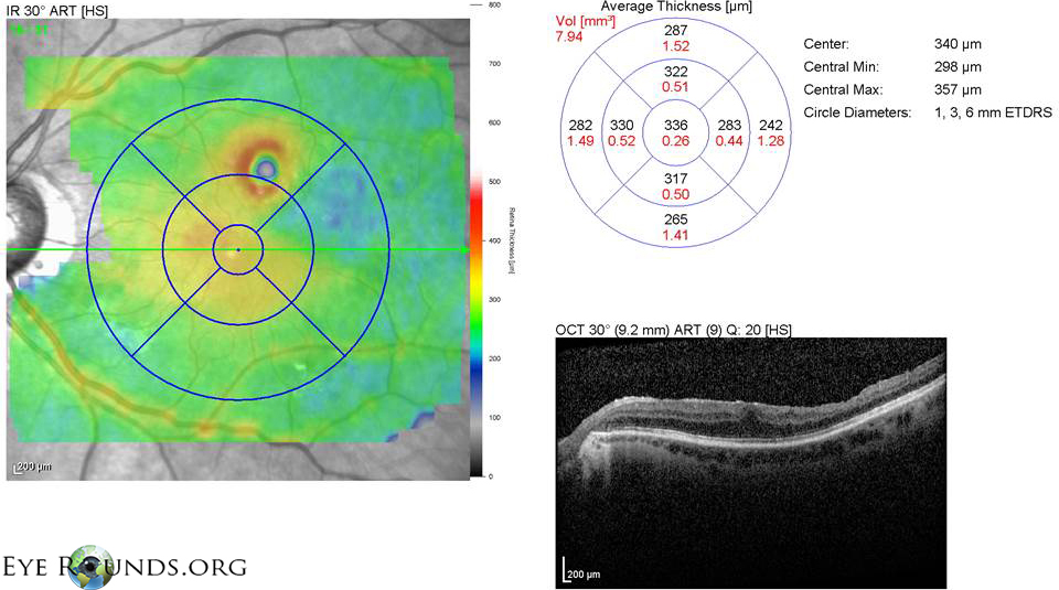
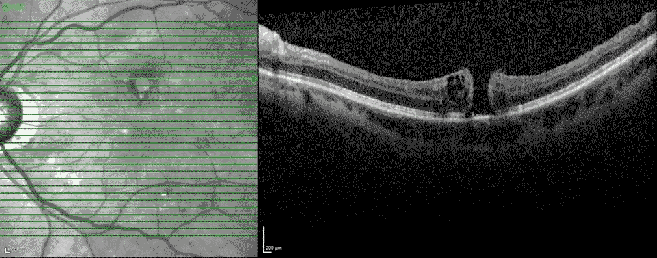


Fig 9: Autofluorescence imaging: hypo and hyper-autofluorescence in areas of pigmentary changes, possibly in areas of forceps grasping during ILM peeling. There is increased-autofluorescence in the area of the eccentric full-thickness macular hole.

Macular Hole, Full Thickness, Extrafoveal/Eccentric
- ILM peeling is sometimes done at the time of macular hole repair, epiretinal membrane removal, and retinal detachment repair.
- Complications can include focal retinal hemorrhages and edema. Direct mechanical trauma can cause nerve fiber layer damage, and iatrogenic eccentric full-thickness retinal breaks and/or holes as shown in this case.
- Paracentral scotomas and visual field defects have also been reported.
References
- Walia HS, Shah GK. ILM peeling a vital intervention for many vitreoretinal disorders. Ophthalmic Surg Lasers Imag 2014;45:92-97.
- Tsuiki E, Fujikawa A, Miyamura N, et al. Visual field defects after macular hole lsurgery with ICG-assisted ILM peeling. Am J Ophthalmol 2007;143:704-5.
- Haritoglou C, Gass CA, Schaumberger M, et al. Macular changes after peeling of the ILM in MH surgery. Am J Ophthalmol 2001;132:363-8.

Ophthalmic Atlas Images by EyeRounds.org, The University of Iowa are licensed under a Creative Commons Attribution-NonCommercial-NoDerivs 3.0 Unported License.