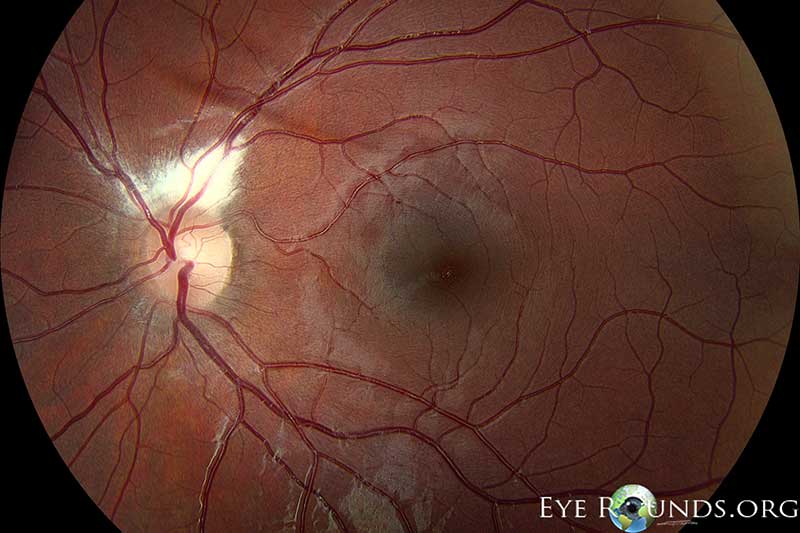
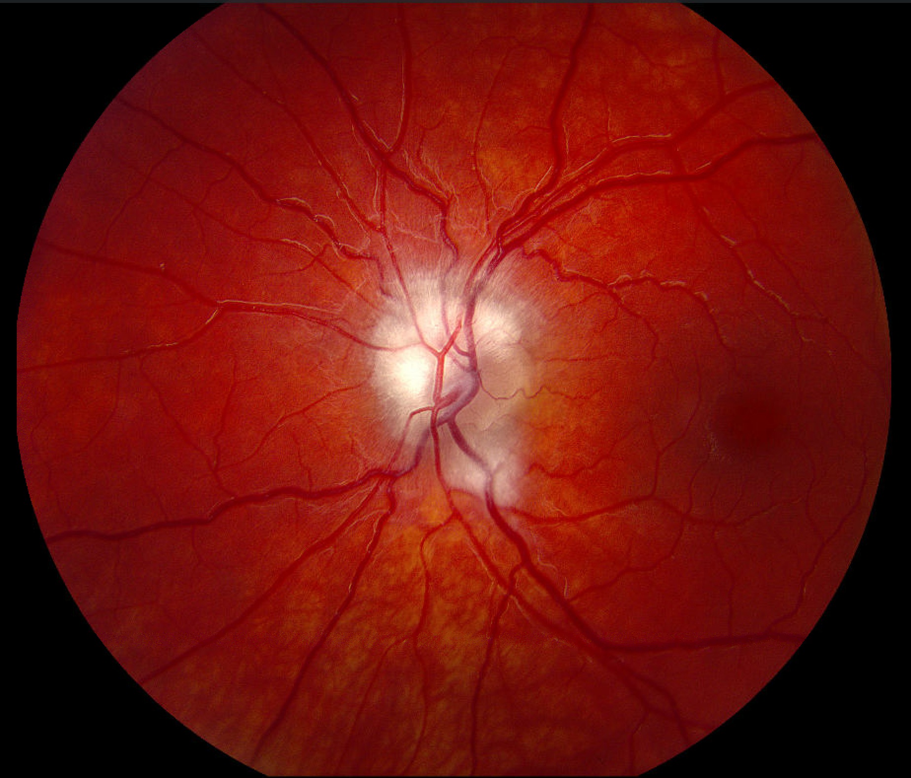
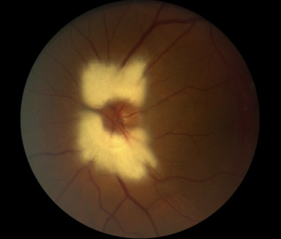
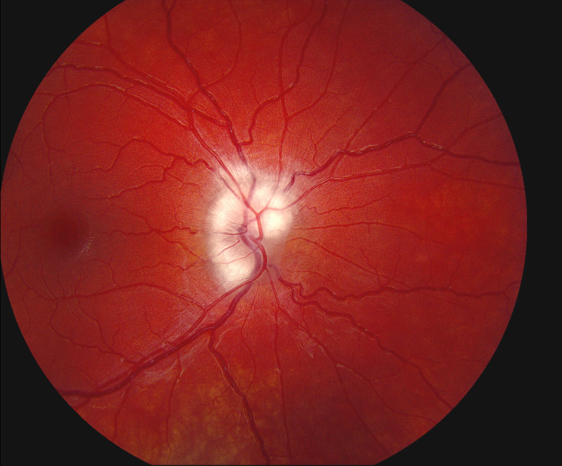
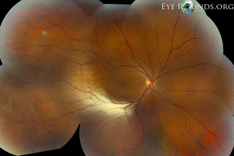
Myelination of the optic nerve fibers normally begins at the lateral geniculate body and ceases at the lamina cribosa. However, in some individuals areas of the retinal nerve fiber layer can also become myelinated. Classically, this finding is unilateral and peripapillary in location, appearing as a white, flame-shaped area with feathered borders as seen in the top photo. However, it can also present bilaterally in 7% of these patients as illustrated in the middle photo.(1) The bottom photo shows a less common presentation affecting a more extensive distribution of the peripheral nerve fibers. Note how the nerve fibers temporal to the macula respect the horizontal raphe, a concept not easily visualized under normal conditions.
Straatsma BR, Foos RY, Heckenlively JR, Taylor GN. Myelinated retinal nerve fibers. American journal of ophthalmology. 1981;91(1):25–38. Available at: http://www.ncbi.nlm.nih.gov/pubmed/7234927.
Vislisel J, Tomlinson LA, Lu O, Chung S, Hackl C, Trejo J, Oetting T. Myelinated Nerve Fiber Layer. EyeRounds.org. Updated March 4, 20026 and August 18, 2025; Original September 9, 2013. Available from https://www.EyeRounds.org/atlas/pages/myelinated-NFL.htm

Ophthalmic Atlas Images by EyeRounds.org, The University of Iowa are licensed under a Creative Commons Attribution-NonCommercial-NoDerivs 3.0 Unported License.


Address
University of IowaLegal
Related Links