

Nodular scleritis: residual scleral thinning
Contributor: William Charles Caccamise, Sr, MD, Retired Clinical Assistant Professor of Ophthalmology, University of Rochester School of Medicine and Dentistry
*Dr. Caccamise has very generously shared his images of patients taken while operating during the "eye season" in rural India as well as those from his private practice during the 1960's and 1970's. Many of his images are significant for their historical perspective and for techniques and conditions seen in settings in undeveloped areas.
Category: External Disease
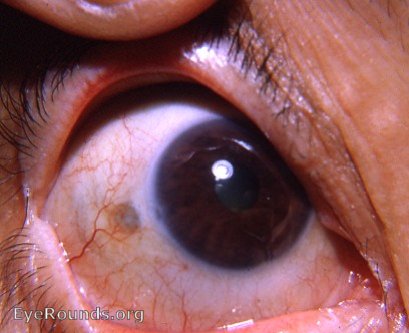
The dark spot on the sclera a few mms from the limbus at 9:00 o'clock is a residual of the nodular scleritis treated by Dr. Caccamise - not a scleral hyaline plaque. The small scar at the limbus at 9:00 o'clock is a residual of a limbal keratitis
Keratitis with scleral thinning from previous nodular scleritis
Both the active keratitis and the previous nodular scleritis are considered to be on an autoimmune basis. The area of scleral thinning is dark because it allows the underlying uveal tissue to be seen.
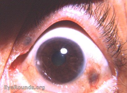
Compare to scleral hyaline plaque - not a residual of nodular scleritis

Ophthalmic Atlas Images by EyeRounds.org, The University of Iowa are licensed under a Creative Commons Attribution-NonCommercial-NoDerivs 3.0 Unported License.