

EyeRounds Online Atlas of Ophthalmology
Contributor: William Charles Caccamise, Sr, MD, Retired Clinical Assistant Professor of Ophthalmology, University of Rochester School of Medicine and Dentistry
*Dr. Caccamise has very generously shared his images of patients taken while operating during the "eye season" in rural India as well as those from his private practice during the 1960's and 1970's. Many of his images are significant for their historical perspective and for techniques and conditions seen in settings in undeveloped areas.
Category: External Disease
Pilosebaceous lid margin cyst
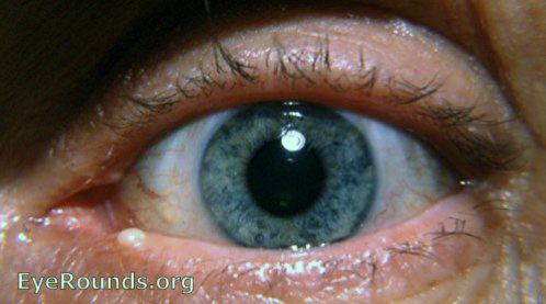
Cysts of the lid margin can be identified basically into to those located anterior to or posterior to the line of separation between the anterior skin-muscle layer and the posterior tarsus-conjunctiva layer. The cyst evident in this photo involves a gland in the anterior lid layer.

Ophthalmic Atlas Images by EyeRounds.org, The University of Iowa are licensed under a Creative Commons Attribution-NonCommercial-NoDerivs 3.0 Unported License.