

EyeRounds Online Atlas of Ophthalmology
Contributor: William Charles Caccamise, Sr, MD, Retired Clinical Assistant Professor of Ophthalmology, University of Rochester School of Medicine and Dentistry
*Dr. Caccamise has very generously shared his images of patients taken while operating during the "eye season" in rural India as well as those from his private practice during the 1960's and 1970's. Many of his images are significant for their historical perspective and for techniques and conditions seen in settings in undeveloped areas.
Category: Cornea
Pseudopterygium
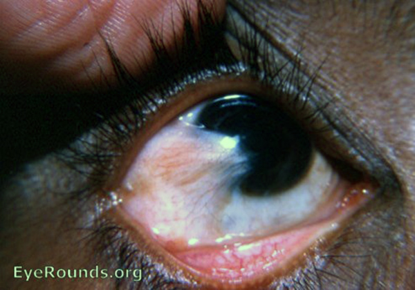
Pseudopterygium is sometimes referred to as cicatricial pterygium. A true pterygium has edges that can be elevated with forceps or under which a probe can be passed A true pterygium aries from a pinguecula. A pseudopterygium arises from destruction of the marginal, corneal epithelium through trauma, e.g. caustics, burns or inflammation.The adjacent conjunctiva migrates to the injured area and becomes fixed to it. A pseudopterygium does not show any tendency to progress. It is able to develop at any point of the corneal circumference.
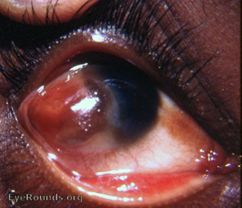
2 pseudopterygia - a small one at 3:30 and a huge one at 8:00 to 9:00.

Ophthalmic Atlas Images by EyeRounds.org, The University of Iowa are licensed under a Creative Commons Attribution-NonCommercial-NoDerivs 3.0 Unported License.