

Laser Refractive Surgery:
From One Medical Student to Another
4. LASIK and PRK
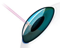
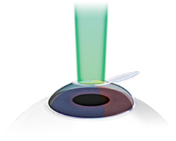
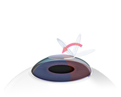
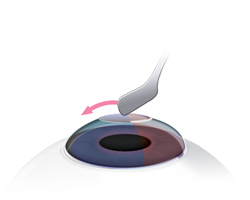
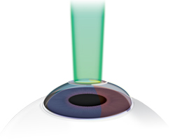
There are two main surgical techniques used to performlaser vision correction: LASIK and PRK. The major difference between these surgeries is the way in which access is gained to the anterior corneal stroma. In LASIK, a 100-200 µm corneal flap is created using a microkeratome (blade) or femtosecond laser (Sakimoto et al. 2006). In PRK, access to the stroma is gained by removing the epithelium of the cornea mechanically (by a brush or blade), chemically (i.e. with alcohol), or by laser (AAO 2007). After stroma is exposed (either by making a flap (LASIK) or by removing the epithelium (PRK)), the patient is asked to fixate on a central light. An eye-tracker is engaged to adjust for any eye movements during the ablation, and a pre-programmed excimer ablation, based on the patient's preoperative refractive error, is performed in seconds to minutes.
Patients are typically given an anxiolytic (i.e. diazepam) prior to surgery to help them relax during the procedure. To decrease infection risk and inflammation, patients generally are treated with an antibiotic drop and steroid drop for about one month after surgery. The patient is examined postoperatively at the following intervals: post-op day 1, 1 week, 1 month, 3 months, 6 months, and 1 year. Illustrations of these surgeries can be seen in Figure 5 and Figure 6.
 |
 |
 |
 |
 |
Item |
LASIK |
PRK |
|---|---|---|
Initial examination |
Similar to PRK |
Similar to LASIK |
Procedural difference |
Corneal flap made with a microkeratome or femtosecond laser |
Surface epithelium removed by a variety of methods (blade, alcohol, brush, or laser) |
Excimer laser procedure |
Similar to PRK |
Similar to LASIK |
Postoperative discomfort |
Usually minimal |
Significant pain for the first several days postoperatively. Patients are given narcotics to control the postoperative pain. |
Visual recovery |
Starts improving at one day |
Starts to improve over the first week, but continues to improve over the first month and may take up to 3 months to achieve the final postoperative visual acuity. |
Possible Complications |
Glare/halo/ghosting |
Glare/halo/ghosting |