

EyeRounds Online Atlas of Ophthalmology
Contributor: William Charles Caccamise, Sr, MD, Retired Clinical Assistant Professor of Ophthalmology, University of Rochester School of Medicine and Dentistry
*Dr. Caccamise has very generously shared his images of patients taken while operating during the "eye season" in rural India as well as those from his private practice during the 1960's and 1970's. Many of his images are significant for their historical perspective and for techniques and conditions seen in settings in undeveloped areas.
Category: Cornea
Bee-sting keratopathy
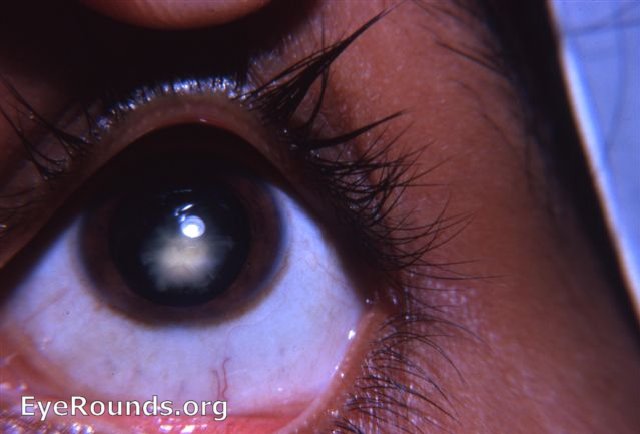
The patient was a young rural patient who came to the Clinic with a complaint of having had a bee sting his eye. Slit-lamp examination revealed a bee-stinger whose tip was in contact with Descemet's membrane. Because of fear of perforating the cornea, the stinger was approached cautiously. However, its barbs prevented removal on the first attempt.The pupil was dilated with atropine. Antibiotic-steroid drops and ointment were provided. The following day, the stinger was successfuly removed. However, the toxins from the stinger combined with the trauma of the surgery led to the leucomatous scarring seen in the photograph. Cases have been reported where a bee-sting has caused uveitis, cataract, and optic neuritis.

Ophthalmic Atlas Images by EyeRounds.org, The University of Iowa are licensed under a Creative Commons Attribution-NonCommercial-NoDerivs 3.0 Unported License.