

Disciform Keratitis due to Herpes Simplex
Contributor: William Charles Caccamise, Sr, MD, Retired Clinical Assistant Professor of Ophthalmology, University of Rochester School of Medicine and Dentistry
*Dr. Caccamise has very generously shared his images of patients taken while operating during the "eye season" in rural India as well as those from his private practice during the 1960's and 1970's. Many of his images are significant for their historical perspective and for techniques and conditions seen in settings in undeveloped areas.
Category: Cornea
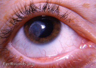
Disciform keratitis due to herpes simplex virus, neovascularized

Fortunately, the vascularized macular scar from the disciform keratitis is not in the visual axis of this eye.

Corneal scar typical of hsv disciform keratitis. Neovascularization is evident.
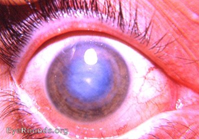
Disciform keratitis (HSV), active with neovascularization

Active neovascularized HSV disciform keratitis.

This cornea is in dire straits. If and when the activity subsides to a sufficient degree, keratoplasty can be attempted. The active neovasculariztion of the entire cornea makes for a very guarded prognosis.
Disciform keratitis due to herpes simplex virus, inactive stage
inactive hsv disciform keratitis
The photo demonstrates an inactive macular scar due to disciform keratitis - a stromal form of herpes simplex virus infection of the cornea.
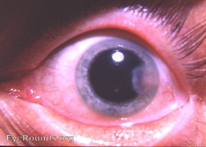
An inactive disciform keratitis scar that unfortunately impinges on the visual axis.

Ophthalmic Atlas Images by EyeRounds.org, The University of Iowa are licensed under a Creative Commons Attribution-NonCommercial-NoDerivs 3.0 Unported License.