

EyeRounds Online Atlas of Ophthalmology
Contributor: William Charles Caccamise, Sr, MD, Retired Clinical Assistant Professor of Ophthalmology, University of Rochester School of Medicine and Dentistry
*Dr. Caccamise has very generously shared his images of patients taken while operating during the "eye season" in rural India as well as those from his private practice during the 1960's and 1970's. Many of his images are significant for their historical perspective and for techniques and conditions seen in settings in undeveloped areas.
Category: External Disease
Lid lesions
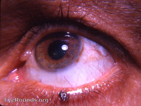
An exact diagnosis clinically of the various palpebral skin lesions by the average ophthalmologist frequently leaves a lot to be desired. Contrariwise, most of these lesions are easily described, categorized, and given a name by the dermatologist who confidently rules supreme in his field of skin esoterica. The lesion seen in the upper lid is a retention cyst of a gland of Moll (see figure 111 in Thiel's Atlas der Augenkrankheiten - probably the leading printed atlas of eye diseases). The pigmented lesion near the lower lid margin is a pigmented papilloma/verruca. It should be excised. There is a danger of malignant degeneration. Conjunctival changes nasal to the limbus at 9 o'clock - seen poorly in the photo - suggest a large pinguecula. Temporal to the limbus at 3 o'clock, there are conjunctival changes that require further evaluation under the slit-lamp. The same applies to the pigment arc overying the limbus in its temporal portion.

Ophthalmic Atlas Images by EyeRounds.org, The University of Iowa are licensed under a Creative Commons Attribution-NonCommercial-NoDerivs 3.0 Unported License.