EyeRounds Online Atlas of Ophthalmology
Contributor: Rekha Khandelwal, MS, DNB, FRCS, Department of Ophthalmology, NKP Salve Institute of Medical Sciences and Lata Mangeshkar Hospital, Nagpur
Category: Cataract
Morgagnian cataract, displaced

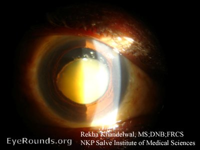
This is a case of bilateral Hypermature morgagnian cataract in a male patient who was 70 years old. Figure 1 (above) shows the right eye with morgagnian cataract and calcified capsule with inferior sinking of the nucleus within the capsular bag.
Interestingly, the patient presented with aphakia in left eye (see Figure 2, velow). There was no history of trauma or surgery done on the eye. On slitlamp examination the calcified posterior capsule is seen just behind the pupillary plane. Indirect ophthalmoscopy revealed the lens nucleus in posterior segment of eye. Due to hard edges of morgagnian cataract, this nucleus likely cut through the thin calcified capsular bag inferiorly and dislocated the lens nucleus into the posterior segmnent.


Ophthalmic Atlas Images by EyeRounds.org, The University of Iowa are licensed under a Creative Commons Attribution-NonCommercial-NoDerivs 3.0 Unported License.


