

EyeRounds Online Atlas of Ophthalmology
Contributor: William Charles Caccamise, Sr, MD, Retired Clinical Professor of Ophthalmology, University of Rochester School of Medicine and Dentistry
*Dr. Caccamise has very generously shared his images of patients taken while operating during the "eye season" in rural India as well as those from his private practice during the 1960's and 1970's. Many of his images are significant for their historical perspective and for techniques and conditions seen in settings in undeveloped areas.
Category: External Disease
Xerotic bulbar conjunctival base of a Bitot's spot
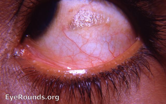
This photograph shows the xerotic patch of bulbar conjunctiva to which the components of a Bitot's spot adhere. Those superficial component's (whipped-up meibomian secretions, conjunctival cellular debris, and bacteria of Corynebacterium xerosis) were removed with a Q-tip. This revealed the underlying xerotic base seen in the photograph. A new, typical foamy Bitot's spot will eventually form in the same area from which the original lesion was removed.

Ophthalmic Atlas Images by EyeRounds.org, The University of Iowa are licensed under a Creative Commons Attribution-NonCommercial-NoDerivs 3.0 Unported License.