

EyeRounds Online Atlas of Ophthalmology
Contributor: William Charles Caccamise, Sr, MD,, Retired Clinical Professor of Ophthalmology, University of Rochester School of Medicine and Dentistry
*Dr. Caccamise has very generously shared his images of patients taken while operating during the "eye season" in rural India as well as those from his private practice during the 1960's and 1970's. Many of his images are significant for their historical perspective and for techniques and conditions seen in settings in undeveloped areas.
Category: Pathology
Anterior ectasiae/Anterior scleral staphlomata
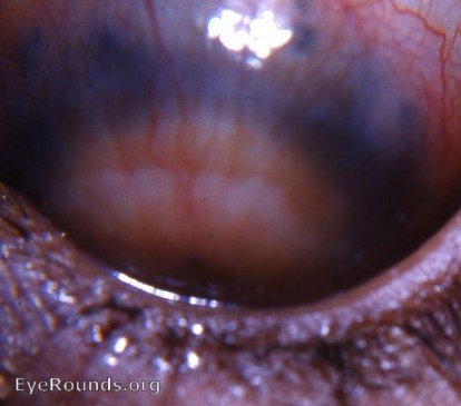
Profound anterior segment pathology frequently follows a perforating corneal ulcer. The photograph presents anterior ectasiae/anterior scleral staphlomata. The differentiation of the ciliary staphyloma seen in the photograph lies between two types,i.e. ciliary staphyloma or intercalary staphyloma. It is frequently difficult to make the exact diagnosis clinically. The pathologist on anatomic study and microscopic study of the enucleated eye will be able to make the exact diagnosis.


Ophthalmic Atlas Images by EyeRounds.org, The University of Iowa are licensed under a Creative Commons Attribution-NonCommercial-NoDerivs 3.0 Unported License.