

EyeRounds Online Atlas of Ophthalmology
Contributor: William Charles Caccamise, Sr, MD, Retired Clinical Assistant Professor of Ophthalmology, University of Rochester School of Medicine and Dentistry
*Dr. Caccamise has very generously shared his images of patients taken while operating during the "eye season" in rural India as well as those from his private practice during the 1960's and 1970's. Many of his images are significant for their historical perspective and for techniques and conditions seen in settings in undeveloped areas.
Atrophia bulbi
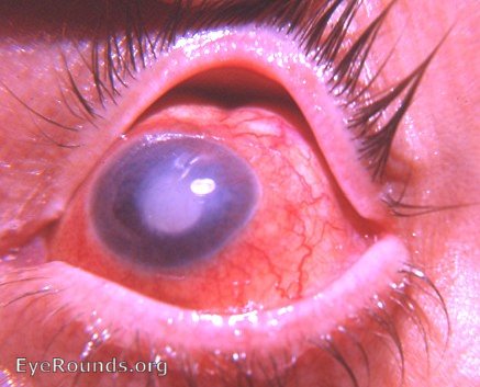
This eye (above) demonstrates the results of longstanding intraocular pathology, e.g. chronic uveitis and retinal detachment. Careful inspection of the photo reveals: hypermature cataracta complicata posterior synechiae extensive neovascularization of the iris and of the pupillary area and the pathognomonic sign of atrophy of the eye - the scleral indentation at 12:30 o'clock.
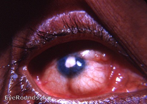
The dense white pupil is due to a cataracta complicata related to longstanding uveitis with longstanding retinal detachment. New bloodvessels can be seen traversing the pupil. The linear indentations in relation to the recti muscles are pathognomonic of shrinkage of the eyeball - atrophia bulbi.
Atrophia bulbi with leukomatous cornea that is showing fatty and calcium infiltration.
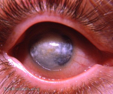
History is of eye problem since early childhood. The cornea is leukomatous with calcium and fatty changes. The eyeball is smaller than normal, i.e. atrophia bulbi due to shrinkage.

Ophthalmic Atlas Images by EyeRounds.org, The University of Iowa are licensed under a Creative Commons Attribution-NonCommercial-NoDerivs 3.0 Unported License.