

EyeRounds Online Atlas of Ophthalmology
Contributor: William Charles Caccamise, Sr, MD, Retired Clinical Assistant Professor of Ophthalmology, University of Rochester School of Medicine and Dentistry
*Dr. Caccamise has very generously shared his images of patients taken while operating during the "eye season" in rural India as well as those from his private practice during the 1960's and 1970's. Many of his images are significant for their historical perspective and for techniques and conditions seen in settings in undeveloped areas.
Category: Cataract
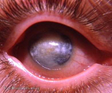
Phthisis bulbi, post cataract surgery

Cataract surgeons must be prepared for potential catastrophic results following their surgery. The photo above is of such a result. The ophthalmologist was a highly skilled, highly experienced and high-volumed cataract surgeon. The eye in the photo above is chronically irritable, sightless, and shrinking. A scleral sulcus is forming along the inferior rectus muscle - a telltale indication of phthisis bulbi. Enucleation was advised.

The linear scleral indentation in line with the tendon of the superiorrectus muscle is pathognomonic of eye shrinkage. Dr. Caccamise as per Dr. Lorenz Zimmerman (USAFI of Pathology) prefers to use the term "atrophia bulbi" for shrinkage of an eye without preceding perforation and "phthisis bulbi" for shrinkage of an eye with preceding perforation.
The linear indentations are always in line with one or more rectus muscles.


Ophthalmic Atlas Images by EyeRounds.org, The University of Iowa are licensed under a Creative Commons Attribution-NonCommercial-NoDerivs 3.0 Unported License.