EyeRounds Online Atlas of Ophthalmology
Contributor: William Charles Caccamise, Sr, MD, Retired Clinical Assistant Professor of Ophthalmology, University of Rochester School of Medicine and Dentistry
*Dr. Caccamise has very generously shared his images of patients taken while operating during the "eye season" in rural India as well as those from his private practice during the 1960's and 1970's. Many of his images are significant for their historical perspective and for techniques and conditions seen in settings in undeveloped areas.
Category: Glaucoma / Iris
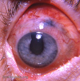
Classic iridencleisis/Holth's operation

Holth's operation/iridencleisis is now a part of abandoned glaucoma surgery. The photo was taken of a right eye that recently had had this procedure for primary open angle glaucoma performed by Dr.Caccamise.In this operation, a limbus based conjunctival flap is dissected. A sclerotomy is performed a short distance from the limbus. The iris is grasped and incised so that one pillar (in this case, the nasal pillar) can be incarcerated through the sclerotomy wound. The conjunctival flap is used to cover the laid out iris and the wound is closed with a running suture. The hoped for result is what is seen in the photo - a visible area of filtration beneath the flap. Notice that only the temporal angle of the iris at the pupil remains - the nasal angle is eliminated when the selected iris pillar has been properly incarcerated in the wound. The fear of sympathetic ophthalmia played a role in the abandonment of this once extremely popular operation for primary open-angle glaucoma.

Ophthalmic Atlas Images by EyeRounds.org, The University of Iowa are licensed under a Creative Commons Attribution-NonCommercial-NoDerivs 3.0 Unported License.


