

Metaherpetic keratitis
Contributor: William Charles Caccamise, Sr, MD, Retired Clinical Assistant Professor of Ophthalmology, University of Rochester School of Medicine and Dentistry
*Dr. Caccamise has very generously shared his images of patients taken while operating during the "eye season" in rural India as well as those from his private practice during the 1960's and 1970's. Many of his images are significant for their historical perspective and for techniques and conditions seen in settings in undeveloped areas.
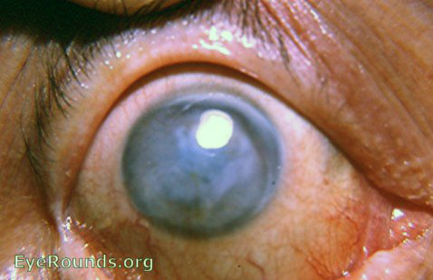
Metaherpetic keratitis is the most severe form of HSV involvement of the cornea. Recurrent attacks of HSV, superficial bullae, stromal involvement, endothelitis, and anterior uveitis can all be part of the metaherpetic phase of HSV. Marked neovascularization is evident in the photo.
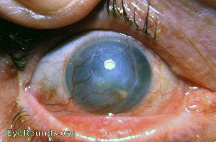
Metaherpetic keratitis shows a widespread stromal involvement of the cornea. Neovascularization is more evident under the slit-lamp. A similar appearing scar but one that is more localized can result from herpetic disciform keratitis.
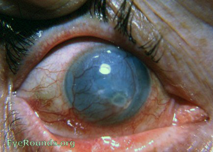
This photo presents catastrophic corneal pathology that started with dendritic keratitis, progressed to disciform keratitis, and ended with highly vascularized and calcifying metaherpetic keratitis. The eye was never a suitable candidate for corneal transplant surgery.

Ophthalmic Atlas Images by EyeRounds.org, The University of Iowa are licensed under a Creative Commons Attribution-NonCommercial-NoDerivs 3.0 Unported License.