

EyeRounds Online Atlas of Ophthalmology
Contributor: William Charles Caccamise, Sr, MD, Retired Clinical Assistant Professor of Ophthalmology, University of Rochester School of Medicine and Dentistry
*Dr. Caccamise has very generously shared his images of patients taken while operating during the "eye season" in rural India as well as those from his private practice during the 1960's and 1970's. Many of his images are significant for their historical perspective and for techniques and conditions seen in settings in undeveloped areas.
Category: Pathology
Scleral hyaline plaque
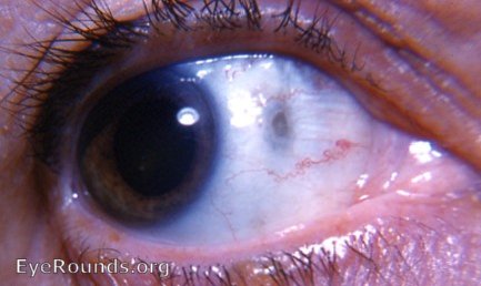
This is a beautiful example of a scleral hyaline plaque. This plaque lies in the palpebral fissure immediately anterior to the medial rectus muscle insertion.The striations in the photo are those of the muscle tendon itself as it inserts into the sclera. The grayish cast to the scleral hyaline plaque is due to the visibilty of the blue uveal tissue through the thinned-out sclera in the scleral hyaline plaque.

Ophthalmic Atlas Images by EyeRounds.org, The University of Iowa are licensed under a Creative Commons Attribution-NonCommercial-NoDerivs 3.0 Unported License.