

EyeRounds Online Atlas of Ophthalmology
Contributor: William Charles Caccamise, Sr, MD, Retired Clinical Assistant Professor of Ophthalmology, University of Rochester School of Medicine and Dentistry
*Dr. Caccamise has very generously shared his images of patients taken while operating during the "eye season" in rural India as well as those from his private practice during the 1960's and 1970's. Many of his images are significant for their historical perspective and for techniques and conditions seen in settings in undeveloped areas.
Category: Trauma
Traumatic cataract with iridodialysis
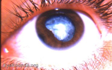
The first photo is computer manipulated to bring out the dark spot of the iridodialysis at 8 o'clock. In untouched photo #2 the iridodialysis area is extremely vague. However, the straight edge of the pupil margin indicates the presence of a dialysis. This is easily verifified by gonioscopy and the slit-lamp examination.
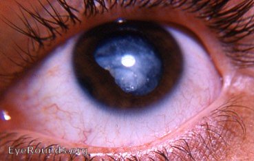
untouched original: iridodialysis seen by slit-lamp and gonioscopy. Straight edge of pupil indicates iridodialysis.
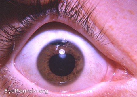
another iridodialysis case
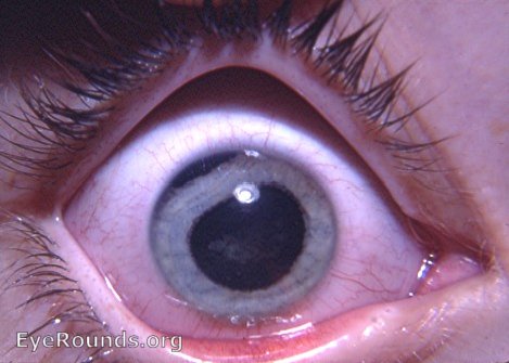
another iridodialysis case

Ophthalmic Atlas Images by EyeRounds.org, The University of Iowa are licensed under a Creative Commons Attribution-NonCommercial-NoDerivs 3.0 Unported License.