

Congenital Ptosis
Congenital fibrosis of the extraocular muscles (CFEOM)
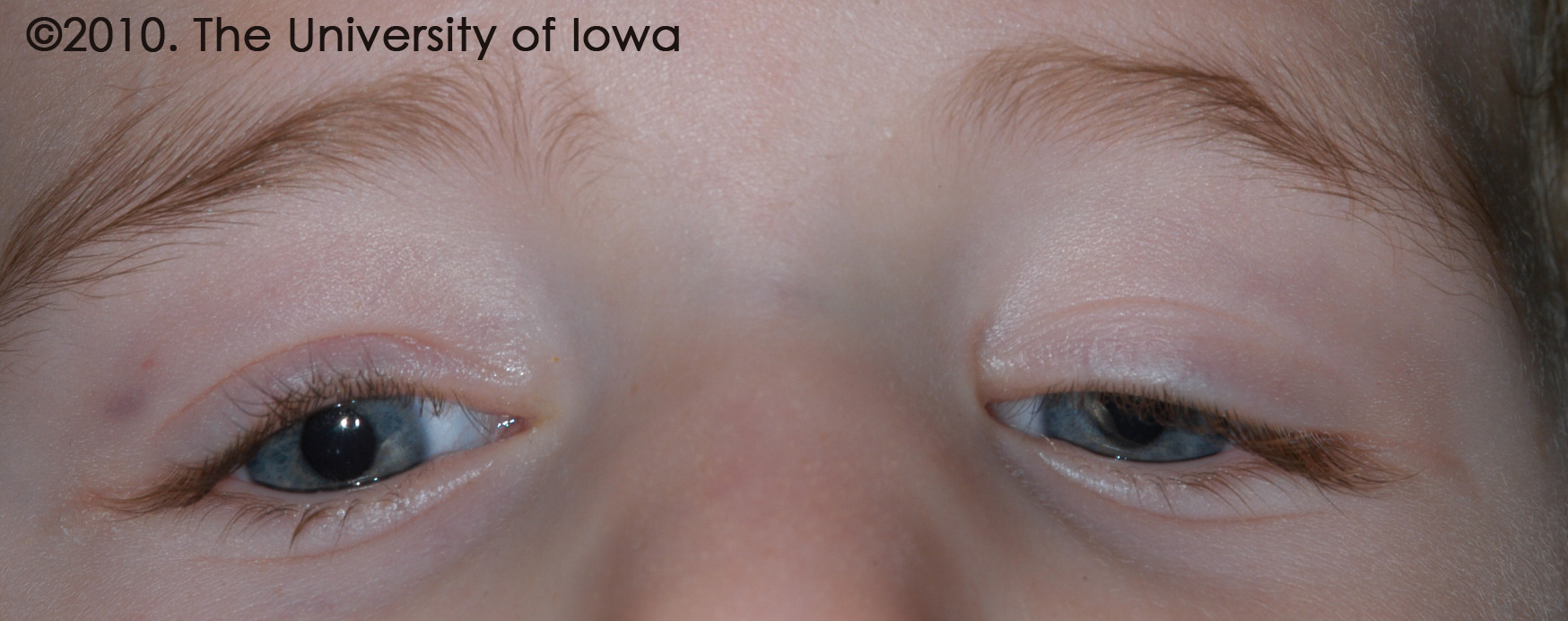
Congenital fibrosis of the extraocular muscles (CFEOM) is a rare, non-progressive condition that results in restrictive global ophthalmoplegia and congenital ptosis. While CFEOM was classically thought to be a myopathy that resulted in muscle fibrosis, there is recent evidence to suggest that the fibrotic changes are secondary to defective innervations of the muscles during development (Heidary 2008). The eyes are most often fixed in an infraducted position, approximately 20-30 degrees below the horizontal midline. This downward gaze, combined with the ptosis, necessitates a chin-up position in many patients (Heidary 2008) (see Figure 6).
Four phenotypes have been described: CFEOM 1, 2, 3 and Tukel syndrome (CFEOM 3 phenotype with postaxial oligodactyly or oligosyndactyly). CFEOM 1 and 3 are inherited in an autosomal dominant fashion and are most commonly associated with missense mutations in the KIF21A gene. While CFEOM 1 typically involves bilateral ptosis and ophthalmoplegia, CFEOM 3 has a more variable phenotype in which ptosis may be unilateral, ophthalmoplegia may be mild and some family members may be unaffected. Mutations in the PHOX2A gene have been associated with CFEOM 2; these are inherited in an autosomal recessive fashion.
These patients require a stepwise surgical approach to correct strabismus and eyelid position. The vertical and horizontal misalignments are addressed first followed by the ptosis repair, as extraocular muscle surgery can alter eyelid position.
 |
BACK NEXT