

Congenital Ptosis
Ptosis Associated with Ocular and Systemic Abnormalities:
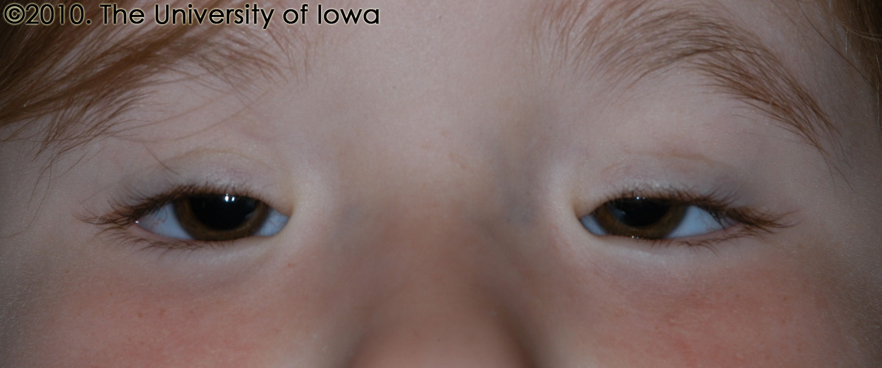
Blepharophimosis-Ptosis-Epicanthus Inversus Syndrome (BPES)
First described by Komoto in 1921, blepharophimosis-ptosis-epicanthus inversus syndrome (BPES) is a dominantly inherited disorder characterized by four features that are present at birth. Patients have:
- severe bilateral, symmetric ptosis,
- telecanthus (an abnormally wide intracanthal distance with normal interpupillary distance)
- epicanthus inversus (skin fold arising from the lower eyelid that covers the medial canthus), and
- blepharophimosis (profound narrowing of the palpebral fissure) (Allen 2008). (See Figure 3.)
The severity of the ptosis and blepharophimosis require most children to adopt a chin-up backwards head-tilt position and to recruit the frontalis in elevating the lids, leading to raised, arched eyebrows. BPES is categorized into two types: Type I is characterized by the abovementioned eyelid findings and premature ovarian failure and infertility. Type II is characterized by eyelid findings without ovarian failure. Other ocular characteristics that have been reported in association with BPES include euryblepharon, strabismus, microphthalmos, lacrimal drainage abnormalities and optic disc coloboma. Extraocular manifestations include a broad, flat nasal bridge, arched palate, and cup-shaped ears (Allen 2008).
BPES is associated with a dominantly inherited mutation in the FOXL2 gene on chromosome 3q23. This gene is expressed primarily in the developing eyelid and in the ovary. Up to 75% of patients with BPES have relatives who have the FOXL2 mutation; the remaining 25% of cases represent either new mutations or milder expression in prior generations (Allen 2008). Type I BPES is characterized by complete penetrance and transmission almost exclusively through males (because of impaired female fertility). Type II BPES is characterized by incomplete penetrance and transmission by both males and females.
The treatment of blepharophimosis requires coordination among oculoplastic surgeons, pediatric ophthalmologists, pediatric endocrinologists and genetic counselors. The surgical repair of the eyelid is complex because of the numerous and interdependent eyelid findings. The repair of ptosis is usually addressed first with frontalis suspension (described later). Early surgery for ptosis is advised if the ptosis is severe and amblyogenic. The canthal repair is usually addressed after ptosis, though some advocate canthal repair first. Medial canthoplasty can be accomplished by a combination of flaps and, at times, transnasal wiring. Some also suggest fixing the medial canthus to the periosteum.
 |